
Glaucoma: A Silent Stealer of Vision
What is Glaucoma?
Today we’re going to discuss glaucoma. Glaucoma is a very serious family of eye diseases. There are many types of glaucoma, but they all have three basic characteristics.
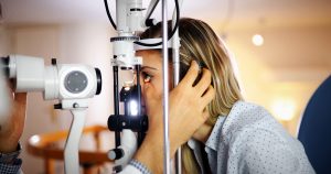
- Intraocular pressure, which causes damage to the nerves in the eye: The eye continuously produces a fluid called the aqueous humor. It circulates within the eye and then drains through a structure called the trabecular meshwork. This fluid produces pressure which fills the eye and gives it its shape, much like air in a tire. Normal pressure is between 10 to 21, but some patients can tolerate higher pressures without apparent damage; this is called ocular hypertension, while others can have damaging effects with normal pressure. This is considered low tension glaucoma. This pressure is easily measured. Most patients have had the experience of the air puff tonometer. A more updated version is called a digital tonometer, which very lightly taps the surface of the cornea with a round, painless probe. This is one of many instruments we can use to measure the intraocular pressure. It is a good screening device, but if you are diagnosed with glaucoma, one of the more accurate instruments are used.
- Changes to the optic nerve in the back of the eye: The optic nerve carries all of the information from the photoreceptors to the brain. If it becomes damaged, the signals traveling through it will become less clear, kind of like having bad reception with an antenna. The optic nerve is visible when we look through the pupil with an instrument called an ophthalmascope. It is a round structure directly in the back of the eye. A healthy nerve looks pink in color and has a flat surface, or perhaps a slight depression called the optic cup. In glaucoma, we see a widening and deepening of the depression and the color changes from light pink to white. If the pressure is not reduced to an acceptable level, this process continues until the entire nerve is affected and can become severely and irreversibly damaged. As we said, the nerve is easily seen through the pupil. Sometimes, we will dilate the pupil and take photos of the nerve to document its appearance and have a reference to compare to in the future as needed. Otherwise, we will just take the photo of the retina if it is clearly visible in the photo and skip the dilation so that your eyes will not need to be blurry and light-sensitive for several hours. There are several other instruments available to measure and evaluate the extent of nerve damage and are used if glaucoma is present.
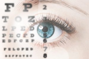
- Peripheral vision loss: When nerve damage has occurred between the eye and the brain, loss of the visual fields may result. Small areas are affected first, in your peripheral vision which the patient doesn’t notice, then larger areas resulting in tunnel vision, and if untreated, eventually total blindness in the eye. Loss of vision in the peripheral fields is the most significant finding in glaucoma and the most diagnostic in relationship to glaucoma. Extensive field loss can be debilitating to the patient, making it hard to perform day to day tasks. Visual fields are measured by instruments called visual field perimeters. They accurately plot a patient’s visual fields and we can see even small defects before they are noticed by the patient. Once plotted, we can use these as a baseline to compare to visual fields taken later. If no progression in field loss is seen we know the patient’s treatment is successful.
Glaucoma is an insidious disease, a silent stealer of vision. The patient has no symptoms, no pain, no visual complaints until the disease has progressed to an advanced state. Early detection is critical in preserving vision because once advanced it is very difficult to prevent further loss. Age, family history of the disease and race are all contributing factors. The mechanisms that cause glaucoma are not known and there is no real cure, only treatment to keep it under control. Treatment usually consists of drops to lower the pressure but some types of glaucoma may require surgical intervention. Yearly eye exams are the best way to catch the disease in an early stage before damage has occurred.
Guest Post Submission from See NC Eyecare. Visit them for all your vision needs in the Raleigh, NC Triangle Area.![]()